Serology: The study of serum or other bodily fluids such as blood, saliva, urine, semen, antibodies, and serum.
|
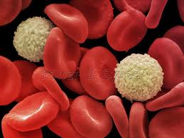
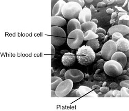
Scanning electron micrograph (SEM) of red blood cells
(erythrocytes, red) and a white blood cell (leucocyte, grey). The disc-shaped red cells transport gases throughout the body. White blood cells are part of the immune system, defending the body against infection. (Spencer, 2012). |
Scanning Electron Microscope
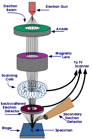
1. A beam of electrons is produced at the top of the microscope by an electron gun.
2. The electron beam follows a vertical path through the microscope, which is held within a vacuum. 3. The beam travels through electromagnetic fields and lenses, which focus the beam down toward the sample. 4. Once the beam hits the sample, electrons and X-rays are ejected from the sample. 5. Detectors collect these X-rays, back-scattered electrons, and secondary electrons and convert them into a signal that is sent to a screen similar to a television screen. This produces the final image. (www.perdue.edu) |
SEM stands for scanning electron microscope.
- uses electrons instead of light to form an image - developed in the early 1950's - has a large depth of field, which allows more of a specimen to be in focus at one time - higher resolution, so closely spaced specimens can be magnified at much higher levels (www.purdue.edu) 
|
|
|
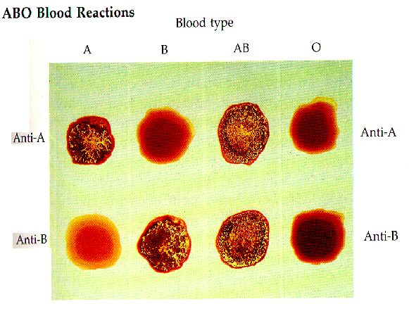
Typing Lab: what type of blood is the following well plate showing?
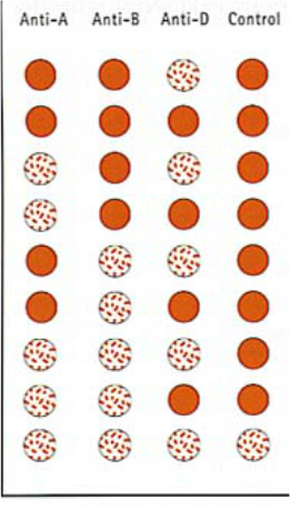
Blood Testing
|
|

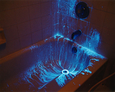
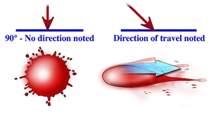
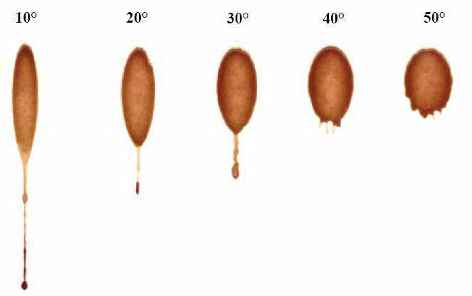
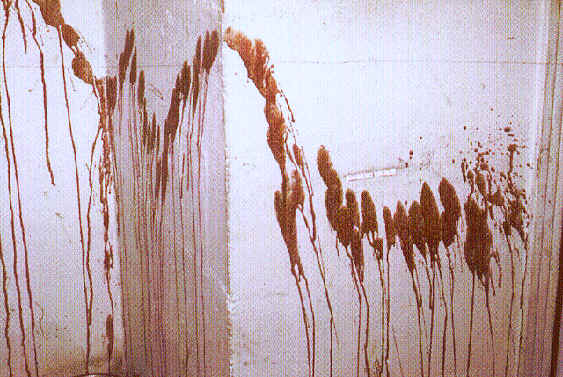
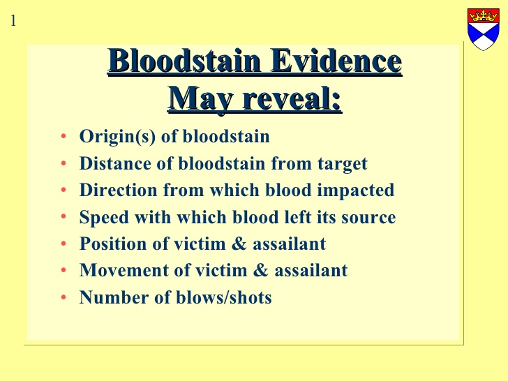
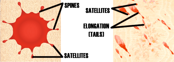
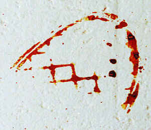
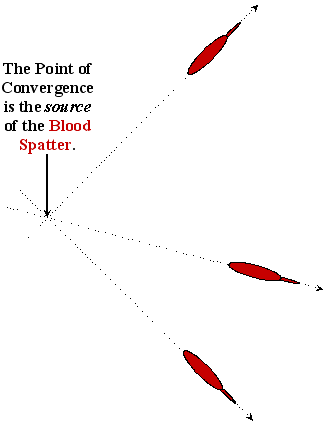
Blood Spatter Analysis
|
Goal- determine the point of origin based on lines of convergence, angle of impact, type of place spatter, and estimated original height.
|

|
|
